Study of Viruses
The study of viruses is known as virology. Viruses can be studied in two ways. The first way is through isolation and cultivation, and the second way through detection, identification and diagnosis. For isolation and cultivation, animals, plants, chick embryo and tissue culture are used. For detection, identification and diagnosis, there are several methods. These methods include tissue culture methods, physical methods, serological methods, immunological methods, and others and molecular biology.
Isolation and Cultivation
Animals and Chick Embryo
Animals and chick embryo were the first method that was used to cultivate virus. This method is rarely used as it is not convenient. However, when preparing for bulk virus, (e.g. antigen or vaccine production) the usage of chick embryo is useful.


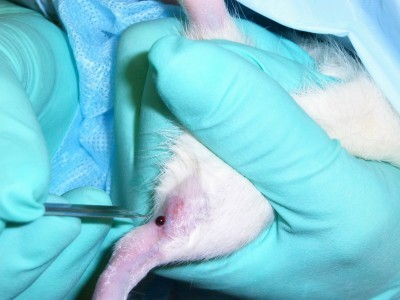
Inoculation of laboratory animals are used when some viruses can only be isolated using this method. Normally, mice are the laboratory animals that are used. Signs of disease or death in animals are observed after inoculation. By testing for neutralization of their pathogenicity for animals by standard sera, the viruses can be identified.


Plants
Tobacco mosaic virus can be found on plants. Virus numbers can be determined by the virus plaques on leaves.

Tissue Culture
The growth of tissue and/or cells that come apart from the organism is known as the tissue culture. There are three types of cell tissue culture – cells grown in vitro, primary cell culture and continuous cell line.
Continuous cell line is obtained from primary cell lines. Continuous cell line can be polyploidy or multiploid.
Detection, Identification and Diagnosis
Tissue Culture Methods
There are two types of tissue culture methods. They are the cytopathic effect (CPE) and the plaque assay.
Cytopathic Effect (CPE)
The degenerative changes of cells that are linked with the multiplication of certain viruses are known as the cytopathic effect (CPE). When in tissue culture, an overlayer of agar restricts the spread of virus. This causes the formation of plaque caused by the cytopathic effect. The examination of the characteristics of cytopathic effect produced on different cell sheets can be used to identify viral infection. However, this technique is not an efficient one, and not all viruses will grow on cell sheets.

Plaque Assay
In 1952, Renato Dulbecco was the first to discover this method. Originally, plaque assay is a virological assay which was introduced to count and measure the infectivity of bacteriaphages. Plaque assay is used to notice and take note of the death of cell in the infected cell culture. When one cell is infected by one virus, it spreads to the surrounding cells. Plaque assay is more accurate when it is at lower concentration.
In 1952, Renato Dulbecco was the first to discover this method. Originally, plaque assay is a virological assay which was introduced to count and measure the infectivity of bacteriaphages. Plaque assay is used to notice and take note of the death of cell in the infected cell culture. When one cell is infected by one virus, it spreads to the surrounding cells. Plaque assay is more accurate when it is at lower concentration.
Plaque assay is very time-consuming and is an easy technique. Plaque assay is only effective for viruses that infect monolayer cells, and for viruses that break down cells. One plaque is formed from one virus on the monolayer. This is the principle of plaque assay.
Plaque assay is used to count only viruses that are capable of multiplying. The culture conditions must be known for virus that is used to study. Samples with very low virus counts use this method. Incubation of virus requires time.
Physical Methods
There are three types of physical methods:
· X-ray crystallography
· Electron microscopy
· Ultracentrifugation
Under electron microscopy, there are transmission electron microscope, scanning electron microscope and STEM. As for ultracentrifugation, one example is the purification.
X-Ray Crystallography
A method that is used to find out the arrangement of atoms within a crystal is known as X-ray crystallography. A beam of X-rays scatters into many directions when it strikes a crystal. According to the angles and intensities of these scattered beams, a three dimensional picture of the density of electrons within a crystal is produced from a crystallography. The mean positions of the atoms in the crystal, their chemical bonding, their disorder and other information can be determined from the electron density.


 X-ray Diffraction Patterns
Electron Microscope
Transmission Electron Microscope
A microscopy method in which a beam of electrons transmit through an ultra thin specimen and interacts with the specimen as they pass through is known as the transmission electron microscopy (TEM). The interaction of the electrons that transmit through the specimen forms an image. The objective lens enlarges and focuses the image onto a fluorescent screen on a layer of photographic film. Fluorescent screen is one of the imaging devices. A sensor can also be used to detect the image. An example of such sensor is the CCD camera.

 Principle features of light and electron microscopes
Scanning Electron Microscope
An electron microscope that images the sample surface by scanning it with a high-energy beam of electrons in a raster scan pattern is known as the scanning electron microscope (SEM). Signals that have information about the sample’s surface topography, composition and other properties such as the ability to conduct electricity are produced when electrons interact with the atoms that make up the sample.
An electron microscope that images the sample surface by scanning it with a high-energy beam of electrons in a raster scan pattern is known as the scanning electron microscope (SEM). Signals that have information about the sample’s surface topography, composition and other properties such as the ability to conduct electricity are produced when electrons interact with the atoms that make up the sample.

Ultracentrifugation
Ultracentrifugation is a process in which a centrifuge spins a rotor at extremely high speeds. Ultracentrifuge has the ability to produce acceleration up to a maximum of 1,000,000 (9,800 km/s²). Purification is one example of ultracentrifugation.


Serological / Immunological Methods
Immunological techniques are used to diagnose virus diseases. This is done so by demonstrating an antigen-antibody reaction to reveal evidence of virus infection. Virus infection can be shown by:
Immunological techniques are used to diagnose virus diseases. This is done so by demonstrating an antigen-antibody reaction to reveal evidence of virus infection. Virus infection can be shown by:
- Serology; where development of antibody to the virus at the time of or just after symptoms of disease.
- Virus being present in the blood of the patients or other tissues.
There are several serological and immunological methods and these methods include:
- Haemagglutination Assay (HA)
- Haemagglutination Inhibition (HI)
- Virus neutralisation
- Complement fixation
- Immunostaining
- Immunofluorescence
- Immuno-Gold Electron Microscope - Immunoprecipitation / Immunoblot
- ELISA
Haemagglutination Assay (HA)
A quantitative of viruses by haemagglutination is known as haemagglutination assay (HA). Haemagglutination is a particular form a agglutination which involves the participation of red blood cells (RBC).
A type of lattice will be formed by red blood cells when viral families with surface or enveloped proteins stick to humans’ or animals’ red blood cells and bind to its N-acetylneuraminic acid.

This method is relatively fast and easy and large amounts of samples use such methods. This method is carried out in the presence of antibody. The type of virus will require different conditions. For example, at certain pH values, some viruses bind RBCs while others bind RBCs at certain ionic strengths.
Haemagglutination Inhibition (HI)
The agglutination of red blood cells by the haemagglutinating viruses are blocked by the antiviral antibody.
Virus Neutralisation
In the case of cell cultures and laboratories animals, antibody prevents or lowers the viral infectivity. This technique is difficult and slow as it requires long and hard work. However, this method is sometimes indispensable.

Complement fixation
Complement fixation test is an immunological medical test, and is used to discover if certain specific antigen or antibody is present in the serum of a patient. This method is widely used to distinguish and find out the cause of infections. It is particularly used in microorganisms that are difficult to identify by culture methods. However, this method is no longer in use in clinical diagnosis as it has already been replaced by serological methods such as ELISA and by DNA-based methods of pathogen detection, especially the polymerase chain reaction (PCR).
Complement fixation test is an immunological medical test, and is used to discover if certain specific antigen or antibody is present in the serum of a patient. This method is widely used to distinguish and find out the cause of infections. It is particularly used in microorganisms that are difficult to identify by culture methods. However, this method is no longer in use in clinical diagnosis as it has already been replaced by serological methods such as ELISA and by DNA-based methods of pathogen detection, especially the polymerase chain reaction (PCR).
In the technique, complement is fixed or used up when antigen reacts with antibody. Complement can be found in guinea pig serum. When red blood cells are added with anti-red-cell antibody, the red cells undergo lyses due to the presence of complement. A positive reaction is indicated by the absence of haemolysis.

Immunostaining
Immunofluorescence
Labeling of antibodies or antigens with fluorescent dyes is known as immunofluoresence. Visualization of the subcellular distribution of biomolecules of interest uses this method. Fluorescence microscope is used to study the immunofluorescent-labelled tissue sections or cultures.

Immuno-gold Electron Microscope
Immuno-gold electron microscope has the same principle as immunofluorescence. The gold particles, measured with a nanometer, are bind to the antibodies. To localize specific proteins or antigens, it is viewed under the electron microscope.

Immunoprecipitation
Immunoprecipitation is a method in which a protein antigen that is precipitated out of solution using an antibody that specifically attaches to that specific protein. This method can be applied when isolating and concentrating a specific protein from a sample that contains many thousands of proteins not of the same kind. Antibody must be coupled to a solid substrate at the same point in the procedure in the technique.

Immunoblot
Immunoblot is also known as Western Blot, and is an analytical method. Detection of particular proteins in a given sample of tissue homogenate or extract uses this method. In this technique, denatured or native proteins, by length of the polypeptide or by 3-D structure of the protein, are separated by the means of gel electrophoresis. After the proteins are transferred to a nitrocellulose membrane, the proteins are then detected using antibodies. Each protein is attached to an antibody, and an antibody is used to detect antigen. A sensitive indicator is used to label an antibody, and there will be a colour reaction with streptavidin.

ELISA
ELISA is also known as the Enzyme-Linked ImmunoSorbent Assay. In immunology, this technique is used to detect whether an antibody or an antigen is present in a sample. The antigen is detected by antibody and an indicator, such as horse radish peroxidise, is used to label the antibody. A colour reaction is shown. In this method, one molecule must be binded to a solid surface.

Molecular Virology
Molecular virology is the study of the organisation of genome, expression of viral genome, replication of genome and the progeny virus.
Molecular Biology and Others
PAGE or SDS PAGE, Western blot, protein sequencing and X-ray crystallography are used to analyse viral proteins. Agarose gels, restriction analysis, sequencing, southern blot, northern blot, PCR o RT-PCR are used to analyse viral genome.
SDS PAGE
SDS-PAGE, sodium dodecyl sulfate polyacrylamide gel electrophoresis, is a method that is commonly used in biochemistry, forensics, genetics and molecular biology. This method is used in the separation of proteins, based on their electrophoretic mobility.


Protein Sequencing
In every cell, there are proteins present. Proteins play an important role in all biological processes. Protein structure is very complicated; hence, to find out a protein structure, first protein sequencing is involved. The first protein sequencing determines the sequences of the constituent peptides of the amino acids and also the conformation it adopts and if it is complex with any non-peptide molecules. Mass spectrometry and Edman degradation reaction are the two major direct methods of proteins sequencing.

Agarose Gels
A technique that is used in biochemistry and molecular to separate DNA or RNA molecules by size is known as the Agarose Gel electrophoresis. To achieve the separation, nucleic acid molecules, with a negative charge, are moved through an agarose mixture with an electrophoresis. Longer molecules move slower and migrate less far than the shorter ones.

Restriction Analysis
A restriction enzyme cuts double-stranded DNA or single-stranded DNA at a particular recognition nucleotide sequences. These particular recognition nucleotide sequences are called the restriction sites.
These enzymes are present bacteria and archaea. These enzymes evolved to give a defense mechanism against viruses that invade them. Inside a bacterial host, the restriction enzymes cut up irrelevant DNA in restriction process. A modification enzyme, methylase, methylates a host DNA to protect it against the activity of restriction enzyme. Restriction modification system is formed from these two processes.

Sequencing
In genetics and biochemistry, seqeuncing refers to the finding of the primary structure or sequence of an unbranched biopolymer. The results of sequencing in a symbolic linear depiction are the sequence which briefly summarizes most of the atomic-level structure of the sequenced molecule. DNA sequencing is one example of sequencing.

Southern Blot
A method that is used in molecular biology to check if a DNA sequence is present in a DNA sample is known as the Southern blot. For size separation of DNA with techniques to transfer the size-separated DNA to filter membrane for probe hybridization, southern blotting is being combined with agarose gel electrophoresis.


Northern blot
A method that is used in molecular biology research to investigate and observe the expression of gene is known as the northern blot. The difference between Northern blot and Southern blot is that Northern blot uses RNA while Southern blot uses DNA. In Northern blot, RNA is analyzed. However, both the Northern blot and the Southern blot use electrophoresis and detection with a hybridization probe.
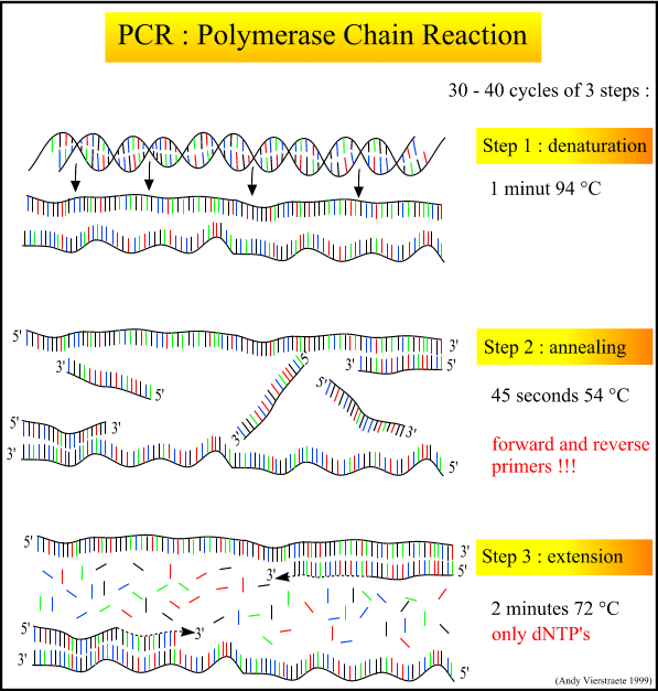
 PCR
PCRPCR refers to polymerase chain reaction. It is a method which is commonly used in molecular biology. The generated DNA is used as a template for replication, as PCR progresses. The DNA template is exponentially amplified when a chain reaction sets in. With PCR, a single or few copies of a piece of DNA across several orders of magnitude can be amplified. Millions or more copies of the DNA piece are generated. A wide array of genetic manipulations is performed by PCR as it can be modified extensively.
Procedures of PCR
PCR normally has a series of 20 to 40 repeated temperature changes and it is known as cycles. Each cycle have 2 to 3 discrete temperature steps. At high temperature, more than 90°C, the cycling is often preceded by a single temperature step. Another single temperature step is followed at the end for the extension of final product or brief storage. The temperatures used and the length of time are dependent on a variety of parameters. These involve the enzyme used for DNA synthesis, concentration of divalent ions and dNTPs in the reaction, and the melting temperature of the primers.
There are six steps in PCR. The six steps are the initialization step, denaturation step, annealing step, elongation step, final elongation step and final hold.
Initialization step: This step involves the heating of reaction to a temperature of 94-96°C and is held for 1 to 9 minutes. However, temperature of 98°C is applied when extremely thermostable polymerases are used. This step only applies to DNA polymerases that need heat activation by hot-start PCR.
Denaturation step: This step is the first regular cycling event and involves the heating of reaction to 94-98°C for 20-30 seconds. DNA template and primers melt as the hydrogen bonds between the complementary bases of the DNA strands are disrupted. Single strands of DNA are then yielded.
Annealing step: This step involves the annealing of the primers to single stranded DNA template, at the temperature of 50 to 65°C for 20 to 40 seconds. When the primer sequence very closely matches the template sequence, stable DNA-DNA hydrogen bonds are formed. DNA synthesis starts when the polymerase attaches to the primer-template hybrid.
Elongated step: Temperature involved in this step is dependent on the DNA polymerase. At this step, a new DNA strand complementary to the DNA template strand is synthesized by the DNA polymerase. This is done so by adding dNTPs that is complementary to the DNA direction. The time that is extended is dependent on both the DNA polymerase that is being used and the legth of the DNA fragment that is to be amplified.
Final elongation step: This step is normally operated at a temperature of 70 to74°C for 5 to 15 minutes after the last PCR cycle. This is to make sure that the remaining single-stranded DNA is extended fully.
Final hold step: When at 4 to15°C, this step, for an indefinite time, can be used for a short-term storage of the reaction.

RT-PCR
RT-PCR refers to reverse transcription polymerase chain reaction. It is used in molecular biology and is a laboratory method for amplifying a defined piece of RNA molecule. DNA complement is formed when the RNA strand is first reverse transcribed. Amplification of the resulting DNA is followed, using polymerase chain reaction.
References
http://www.ogpbb.com/accessories/incubation-guide/images/chicken-embryo-development.jpg
http://news.bbc.co.uk/olmedia/185000/images/_189098_chick_egg_150.jpg
http://wwwdelivery.superstock.com/WI/223/1597/PreviewComp/SuperStock_1597-11330.jpg
http://www.medipoint.com/assets/images/Rat_-_Bloodcollectionaftersaphenousbleed.JPG
http://www.apsnet.org/online/feature/tobacco/image/tobacco1.jpg
http://en.wikipedia.org/wiki/Tissue_culture
http://en.wikipedia.org/wiki/Cytopathic_effect
http://www.scielosp.org/img/revistas/rsp/v34n4/2532f1.jpg
http://www.biology-online.org/dictionary/Plaque_assay
http://en.wikipedia.org/wiki/X-ray_crystallography
http://www3.imperial.ac.uk/pls/portallive/docs/1/872005.JPG
http://campus.queens.edu/faculty/jannr/Genetics/images/bx13_01a.jpg
http://research.yale.edu/ysm/images/77.2/articles-protein-xray.jpg
http://en.wikipedia.org/wiki/Transmission_electron_microscope
http://www.nims.go.jp/htm21/MA/tem.jpg
http://www.mih.unibas.ch/Booklet/Lecture/Chapter1/Fig.1-6.gif
http://www.microscopehelp.com/images/06.jpg
http://lcac1.loras.edu/che/Ultracentrifuge.jpg
http://www.uklabs-direct.co.uk/assets/applets/Sorval_RT_Legend_Ref_Centrifuge__1.JPG
http://en.wikipedia.org/wiki/Hemagglutination
http://en.wikipedia.org/wiki/Hemagglutination_assay
http://upload.wikimedia.org/wikipedia/en/1/17/Anti-HLA_agglutinated_RBC.PNG
http://www.biobest.co.uk/images/vnt.jpg
http://en.wikipedia.org/wiki/Complement_fixation_test
http://www.dshs.state.tx.us/LAB/images/cf_test2.gif
http://en.wikipedia.org/wiki/Immunofluorescence
http://www.abcam.com/ps/datasheet/images/ab3298_12.JPG
http://www.nature.com/emboj/journal/v20/n9/images/7593720f3.jpg
http://en.wikipedia.org/wiki/Chromatin_immunoprecipitation#Chromatin_Immunoprecipitation_.28ChIP.29
http://www.molecularstation.com/images/immunoprecipitation.gif
http://en.wikipedia.org/wiki/Western_blot
http://www.eurogentec.com/EGT/images/Eurogentec-proteomics-identification-western-blot-530x407.jpg
http://en.wikipedia.org/wiki/ELISA
http://64.202.120.86/upload/image/articles/2006/biopen/biopen-elisa-schematic.jpg
http://en.wikipedia.org/wiki/SDS-PAGE
http://www.steve.gb.com/images/science/sds_page.png
http://www.adtrendering.com.au/_resource/image/sds-page.jpg
http://www.wiley.com/legacy/college/boyer/0470003790/cutting_edge/shotgun_seq/bioinformatics.jpg
http://en.wikipedia.org/wiki/Protein_sequencing
http://en.wikipedia.org/wiki/Agarose_gel_electrophoresis
http://www.molecularstation.com/images/agarose-gel-electrophoresis.jpg
http://en.wikipedia.org/wiki/Restriction_enzyme
http://homepages.strath.ac.uk/~dfs99109/BB211/Lod7-5bMethyl.JPG
http://en.wikipedia.org/wiki/Sequencing
http://www.nucleics.com.au/images/site_images/uniseq-dna-sequencing-principle.gif
http://en.wikipedia.org/wiki/Southern_blot
http://www.molecularstation.com/images/southern-blot.jpg
http://www.mun.ca/biology/scarr/Southern_autoradiogram_with_RFLP.gif
http://en.wikipedia.org/wiki/Northern_blot
http://www.biochemsoctrans.org/bst/031/0781/bst0310781f03.gif
http://en.wikipedia.org/wiki/Polymerase_chain_reaction
http://en.wikipedia.org/wiki/RT-PCR
http://users.ugent.be/~avierstr/principles/pcrsteps.gif
8 comments:
great thanks
This is a very remarkable job well done. It is very resourceful.
More grease to ur elbows
Thanks for shearing about this I thinks its very hopeful post and very important post for us.thanks for your great and helpful presentation I like your good service.I always appreciate your post.
assay sample
Professionally written blogs are rare to find, however I appreciate all the points mentioned here. I also want to include some other writing skills which everyone must aware of.
ติว sat ที่ไหน ดี
Fantastic post, very informative. I wonder why the other specialists of this sector do not notice this. You must continue your writing. I'm confident, you have a great readers' base already!
ผ้าห่มขนหนู
Investing online has been a main source of income,that's why knowledge plays a very important role in humanity,you don't need to over work yourself for money.All you need is the right information,and you could build your own wealth from the comfort of your home!Binary trading is dependent on timely signals,assets or controlled strategies which when mastered increases chance of winning up to 90%-100% with trading. It’s possible to earn $10,000 to $20,000 trading weekly-monthly,just file a complaint with Robert,I had almost given up on everything about binary trading and ever getting my lost funds back,till i met with him,with his help now i have my lost funds back to my bank account and I can now trade successfully with his profitable strategies and software!! Email: Robertseaman939@gmail.com or Fb.me/investandmakemoney1 or whatsApp: +44 7466 770724
I'm 61 years old, I contracted hpv in 2011' I has be taking lot treatment for it and embarrassed some months ago the wart stated coming out seriously, I used lot recommendation because there was lot warts around my anus and was so . but today I'm totally happy I got the virus eliminated by using natural treatment from Dr Onokun herbal center after his treatment I got cured. all the warts went away' seriously believed Dr Onokun he have the cure for human papillomavirus because he has eliminated hpv been in my body since 2011, Dr Onokun make it possible for me. Here is Dr Onokun email: dronokunherbalcure@gmail.com or website: https:// Dronokunherbalcure.wordpress.com/ page at: https://www.facebook.com/naturaltreatment1
I was diagnosed as HEPATITIS B carrier in 2013 with fibrosis of the
liver already present. I started on antiviral medications which
reduced the viral load initially. After a couple of years the virus
became resistant. I started on HEPATITIS B Herbal treatment from
ULTIMATE LIFE CLINIC (www.ultimatelifeclinic.com) in March, 2020. Their
treatment totally reversed the virus. I did another blood test after
the 6 months long treatment and tested negative to the virus. Amazing
treatment! This treatment is a breakthrough for all HBV carriers.
Post a Comment